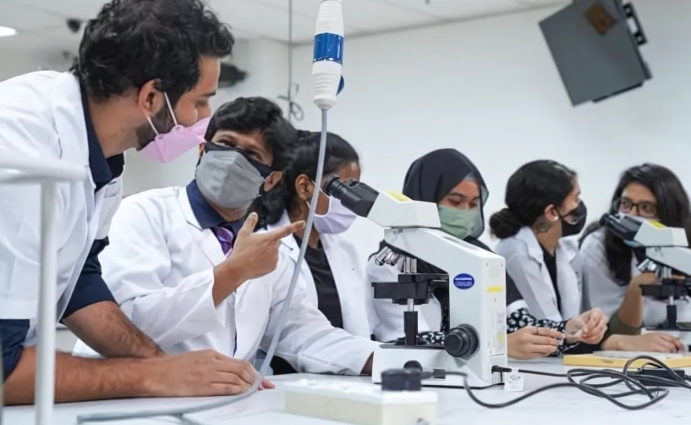
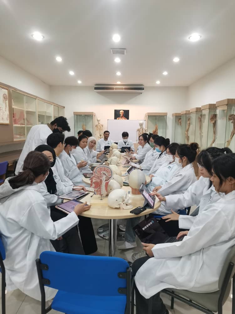
At SEGi University’s Faculty of Medicine, learning extends far beyond lectures and textbooks. One of our most valuable assets is the Anatomy and Pathology Museum, along with the Anatomy Preparation Room that bring the intricacies of the human body to life in a tangible and impactful way.
The museum showcases an extensive collection of preserved specimens, anatomical models, and interactive exhibits covering all major body systems. A standout feature is our cadaver-based learning experience, which enables students to explore real human anatomy in exceptional detail. SEGi is among the few institutions in the country that offers hands-on cadaveric dissection training as early as the first year, a practice that significantly enhances surgical understanding during clinical training.
In addition to anatomy, the museum includes a wide array of pathology specimens that illustrate how various diseases affect organs and tissues. By examining both healthy and diseased samples side by side, students sharpen their diagnostic skills and gain a deeper insight into pathological knowledge that proves invaluable during their clinical years.
Integrated from the early stages of the MBBS curriculum, the museum supports both pre-clinical and clinical education. Students first establish a solid foundation in anatomy and then revisit these resources to reinforce their learning before starting hospital rotations.
With comprehensive facilities and support from experienced lecturers, medical scientists, and clinical consultants, we are committed to providing our students with the best possible learning environment. This immersive training equips them with the confidence and competence to thrive in real-world medical practice.
Discover more about SEGi’s MBBS programme here and how our exceptional facilities give future doctors a head start in their medical careers.
About the author:
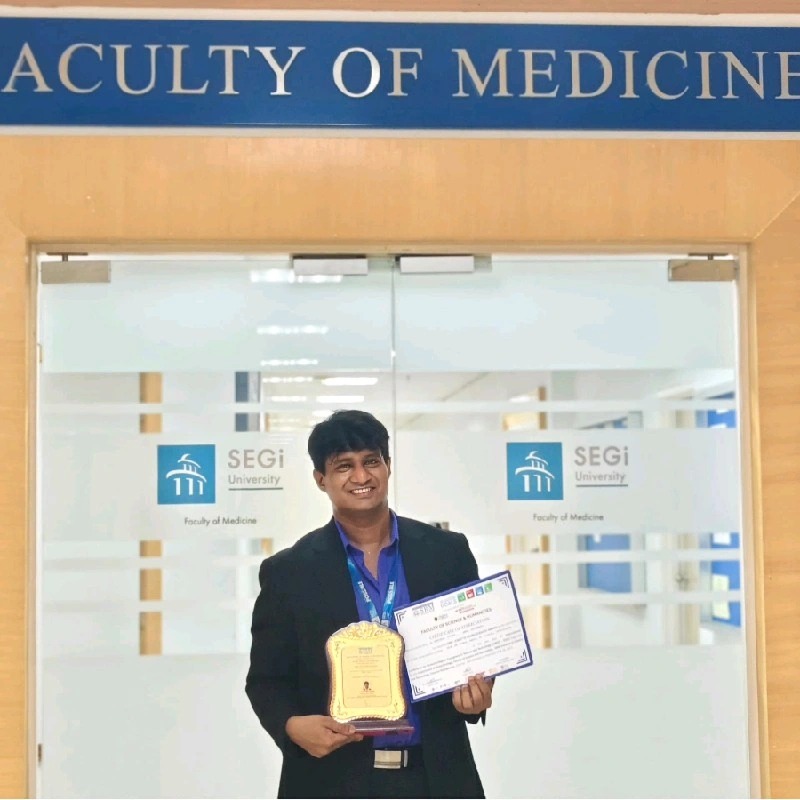
ASSOC. PROF. DR. SARAVANAKUMAR (SK)
Associate Professor of Anatomy, Faculty of Medicine, SEGi Universty.
Assoc. Prof. Dr. Saravanakumar is a highly accomplished dental surgeon with a double master’s degree in Medical Anatomy and Psychology, and a PhD in Medical Anatomy focusing on stem cell regeneration. A member of the Royal College of Surgeons (UK) and the British Association of Clinical Anatomists (UK), as well as a registered clinical practitioner with the Dental Council, he has over 15 years of experience teaching anatomy to medical and dental students. His expertise spans gross anatomy, histology, embryology, neuroanatomy, and osteology, complemented by an impressive portfolio of more than 50 published research articles, a patent, and a government-funded research grant on mesenchymal stem cells. Recognised with multiple awards for research, teaching, and professional excellence, he has served as an examiner at various academic levels and as a keynote speaker and chairperson at national and international conferences, including the Malaysian Surgical Society.